Ventricular Tachycardia: Cellular Electrophysiology, Molecular Mechanisms and Arrhythmogenic Substrates
Explore the molecular and cellular biology of ventricular tachycardia, including ion channels, calcium handling, myocardial scar, re-entry, oxidative stress and inherited channelopathies.
1. What is ventricular tachycardia?

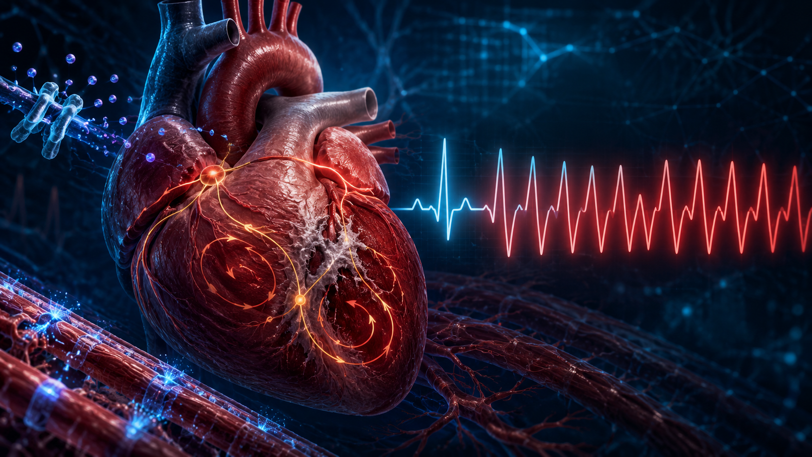
Ventricular tachycardia, commonly abbreviated as VT, V tach or VTach, is a sequence of rapid electrical activations originating below the atrioventricular node. The source may be ventricular myocardium, scar-border tissue, the ventricular outflow tract or the specialised His–Purkinje conduction system.
VT is not one single biological disorder. It represents a final electrical phenotype that can emerge from several molecular and tissue-level mechanisms. The principal mechanisms are:
Re-entry around or through an anatomical substrate
Triggered activity caused by afterdepolarisations
Abnormal or enhanced automaticity
The interaction between a trigger and a susceptible myocardial substrate determines whether an isolated ventricular beat terminates or develops into sustained ventricular tachycardia.
2. Normal ventricular electrical biology
Understanding ventricular tachycardia begins with the normal ventricular action potential.
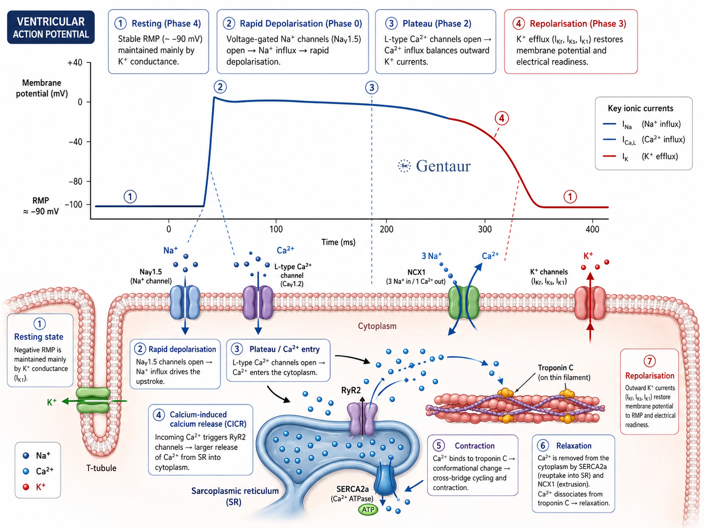
A ventricular cardiomyocyte maintains a negative resting membrane potential through potassium-selective conductance. When an electrical wave reaches the cell, voltage-gated sodium channels open rapidly. The resulting inward sodium current, principally mediated by Nav1.5, produces rapid membrane depolarisation.
The action-potential plateau is supported by calcium entry through L-type calcium channels. This calcium influx activates ryanodine receptor type 2 channels, or RyR2, in the sarcoplasmic reticulum. RyR2 releases a larger quantity of stored calcium into the cytoplasm, initiating myofilament contraction through calcium binding to troponin C.
Relaxation requires calcium removal from the cytoplasm. Calcium is pumped back into the sarcoplasmic reticulum by SERCA2a and extruded from the cell primarily by the sodium-calcium exchanger, NCX1. Potassium currents subsequently repolarise the membrane and restore electrical readiness for the next activation.
| Biological component | Normal function | Arrhythmogenic consequence of dysfunction |
|---|---|---|
| Item one | Description | Value |
| Nav1.5 sodium channel | Rapid ventricular depolarisation | Reduced excitability, slowed conduction or abnormal persistent sodium current |
| L-type calcium channel | Initiates calcium-induced calcium release | Excess inward current and prolonged action potentials |
| RyR2 | Releases calcium from the sarcoplasmic reticulum | Diastolic calcium leak and delayed afterdepolarisations |
| SERCA2a | Returns cytosolic calcium to the sarcoplasmic reticulum | Disturbed calcium loading and relaxation |
| NCX1 | Extrudes calcium in exchange for sodium | Transient inward current following spontaneous calcium release |
| Potassium channels | Repolarise the action potential | Prolonged or abbreviated refractoriness |
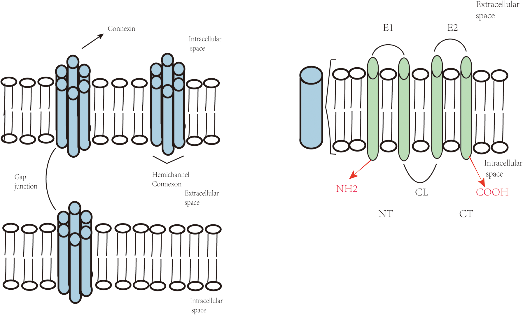
| Connexin 43 | Electrically couples adjacent cardiomyocytes | Conduction slowing and increased electrical heterogeneity |
3. From a single cardiomyocyte to ventricular tachycardia
A molecular defect in one cardiomyocyte is not always sufficient to generate sustained VT. The abnormal electrical event must overcome electrotonic suppression by neighbouring cells and propagate through enough myocardium to recruit a larger ventricular circuit.
Ventricular arrhythmogenesis is therefore a multiscale process:
Molecular abnormality ↓
Ion-channel or calcium-handling dysfunction
↓
Abnormal cardiomyocyte membrane potential
↓
Triggered ventricular activation
↓
Propagation through a vulnerable tissue substrate
↓
Sustained ventricular tachycardiaThis multiscale framework explains why an identical molecular abnormality may produce no arrhythmia in one situation but sustained VT during ischaemia, sympathetic stimulation, electrolyte disturbance or structural remodelling.
4. Scar-mediated re-entry in monomorphic ventricular tachycardia
4.1 Formation of the arrhythmogenic scar
After myocardial infarction, cardiomyocyte death is followed by inflammation, extracellular-matrix deposition and replacement fibrosis. Mature scar tissue is largely non-excitable, but the scar is rarely electrically uniform.
Surviving cardiomyocyte bundles may remain within or along the border of fibrotic tissue. These myocardial strands can be separated by collagen and may form narrow pathways through which electrical activation travels slowly and discontinuously. Histological and electrophysiological studies of human post-infarction hearts established that such surviving myocardial pathways can form the anatomical basis of re-entrant VT.
4.2 The re-entry circuit
Re-entry requires:
A pathway capable of conducting an electrical impulse
Unidirectional conduction block
Sufficiently slow conduction
Recovery of excitability before the impulse returns
An impulse entering the scar border may be blocked in one direction while proceeding slowly through another channel. By the time it exits and returns to the original tissue, that tissue may have recovered excitability. The wave can then re-enter the pathway and circulate repeatedly.
The narrow surviving myocardial region essential for maintaining this circuit is often described as the critical isthmus. Because successive ventricular activations follow a similar circuit, the resulting QRS complexes tend to maintain a consistent morphology, producing monomorphic ventricular tachycardia..
4.3 Cellular changes inside the scar border zone
Slow conduction is not caused by collagen alone. Cardiomyocytes in the infarct border zone can undergo extensive electrical remodelling, including:
Reduced sodium-current availability
Altered sodium-channel localisation
Reduced gap-junction density
Lateral redistribution of connexin 43
Abnormal cell geometry
Increased extracellular resistance
Heterogeneous action-potential duration
Sympathetic nerve remodelling
The combination of reduced excitability and disrupted cell-to-cell coupling causes electrical activation to follow a slow zigzag path through surviving myocardial bundles.
5. Gap junctions, connexin 43 and conduction heterogeneity
6. Triggered activity and abnormal calcium biology
7. Catecholaminergic polymorphic ventricular tachycardia
Catecholaminergic polymorphic ventricular tachycardia, or CPVT, illustrates how disturbed calcium biology can cause severe ventricular arrhythmias in a structurally normal heart.
CPVT is commonly associated with pathogenic variants in:
RYR2, encoding the cardiac ryanodine receptorCASQ2, encoding calsequestrin-2Less commonly, other calcium-regulatory genes
RyR2 variants frequently increase the probability of inappropriate calcium release from the sarcoplasmic reticulum. CASQ2 variants disrupt calcium storage and regulation within the sarcoplasmic reticulum.
8. Polymorphic ventricular tachycardia and torsades de pointes
Polymorphic ventricular tachycardia is characterised by continuously changing QRS morphology. Biologically, the changing ECG pattern reflects unstable or shifting ventricular activation pathways rather than repeated activation through one fixed circuit.
Torsades de pointes is a specific form of polymorphic ventricular tachycardia associated with delayed ventricular repolarisation and QT prolongation.
At the molecular level, torsades susceptibility is influenced by the balance between inward and outward currents during the action-potential plateau. Reduced repolarising potassium currents or increased inward sodium and calcium currents prolong action-potential duration.
The risk is not determined by QT duration alone. Spatial and temporal heterogeneity of repolarisation, pause dependency, early afterdepolarisations and the availability of vulnerable ventricular tissue also contribute to arrhythmia initiation.
Repolarisation reserve
The concept of repolarisation reserve describes the myocardium’s ability to maintain stable repolarisation when one repolarising mechanism is impaired.
A person may tolerate partial inhibition of one potassium current because other currents compensate. However, the addition of hypokalaemia, bradycardia, structural disease, genetic susceptibility or a second medication may exhaust this reserve and allow marked action-potential prolongation.
This biological model helps explain why only a subset of patients exposed to a QT-prolonging drug develop torsades de pointes.
9. The His–Purkinje system as an arrhythmogenic structure
The His-Purkinje system rapidly distributes electrical activation throughout the ventricles. Purkinje cells differ biologically from working ventricular cardiomyocytes in their ion-channel expression, conduction properties, action-potential duration and calcium handling.
Purkinje tissue can :
Generate focal ventricular activity
Form part of macro-re-entrant circuits
Interact with myocardial scar
Initiate ventricular fibrillation
Participate in bundle-branch re-entry
In structural heart disease, the Purkinje-myocardial junction may become particularly arrhythmogenic. Surviving Purkinje fibres near infarcted myocardium can provide triggers or form part of the VT circuit.
10. Heart failure and arrhythmogenic remodelling
11. Mitochondria, oxidative stress and ventricular arrhythmogenesis
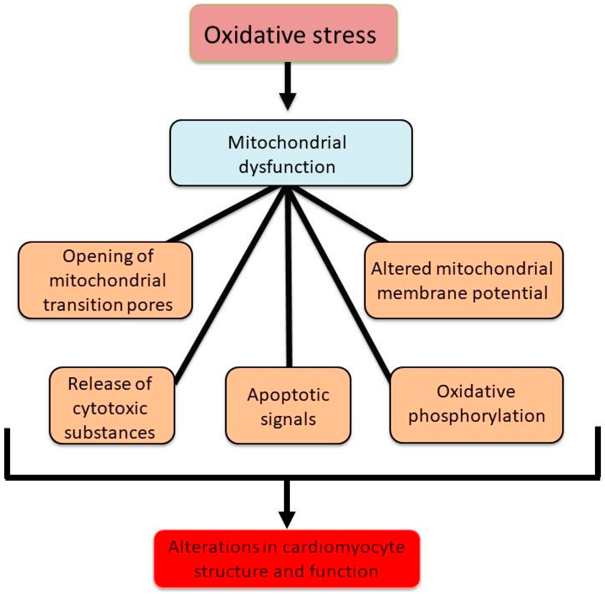
Mitochondria supply ATP required for contraction, ion transport and restoration of ionic gradients. They also regulate calcium signalling, redox balance and cell survival.
Mitochondrial dysfunction can promote arrhythmias through:
Reduced ATP production
Excess reactive oxygen species
Abnormal mitochondrial calcium uptake
Impaired sodium and calcium homeostasis
Oxidation of RyR2
Modification of Nav1.5
Activation of ATP-sensitive potassium channels
Gap-junction remodelling
Reactive oxygen species can oxidise RyR2 and increase diastolic calcium leak. They may also modify sodium channels, calcium-handling proteins and connexins. These effects simultaneously increase triggered activity and reduce conduction stability.
Mitochondrial calcium is normally used to match ATP production with cardiac workload. Excessive mitochondrial calcium, however, can increase oxidative stress and contribute to mitochondrial permeability transition and cardiomyocyte injury. Conversely, inadequate mitochondrial calcium uptake may limit ATP production during increased workload.
The relationship is therefore bidirectional: calcium instability damages mitochondrial function, while mitochondrial dysfunction further destabilises calcium cycling and membrane electrophysiology.
13. Biological rationale of ventricular tachycardia treatments
Frequently asked scientific questions
What is the main cellular mechanism of ventricular tachycardia?+
There is no single mechanism. Scar-related monomorphic VT is commonly maintained by re-entry, while some idiopathic and inherited forms arise from triggered activity caused by abnormal calcium cycling or afterdepolarisations.
How does myocardial fibrosis cause ventricular tachycardia?+
Fibrosis separates surviving cardiomyocytes, disrupts gap-junction coupling and creates spatially heterogeneous conduction. Narrow pathways of viable myocardium within scar tissue can conduct slowly and form re-entrant circuits.
How does RyR2 dysfunction cause ventricular tachycardia?+
Abnormal RyR2 opening allows spontaneous calcium release from the sarcoplasmic reticulum during diastole. NCX1 responds by extruding calcium and generating an inward current, which can produce delayed afterdepolarisations and triggered ventricular beats.
What is the biological difference between monomorphic and polymorphic VT?+
Monomorphic VT usually reflects repeated ventricular activation through one relatively stable focus or circuit. Polymorphic VT reflects continuously changing activation pathways, commonly associated with repolarisation instability, acute ischaemia or abnormal intracellular calcium release.
Can ventricular tachycardia occur without structural heart disease?+
Yes. Inherited channelopathies and calcium-handling disorders can produce VT in a heart without visible structural abnormalities. CPVT is a major example and is commonly associated with RYR2 or CASQ2 dysfunction.
Why can ventricular tachycardia reduce blood pressure?+
Rapid ventricular activation shortens filling time and may produce mechanically uncoordinated contraction. Stroke volume falls, reducing cardiac output. The haemodynamic effect depends on ventricular rate, ventricular function, activation pattern and the presence of structural heart disease.
“Ventricular tachycardia should be understood as a biological systems disorder rather than only an ECG abnormality. At the molecular level, changes in sodium, calcium and potassium currents alter excitability and repolarisation. Abnormal RyR2 activity and calcium cycling generate afterdepolarisations. Mitochondrial dysfunction and reactive oxygen species modify ion channels, calcium-regulatory proteins and gap junctions. At the tissue level, fibrosis, infarct scar and connexin remodelling create regions of conduction block and slow propagation. These regions can transform a transient ventricular trigger into a sustained re-entrant rhythm. The resulting ECG phenotype monomorphic VT, polymorphic VT, torsades de pointes or bidirectional VT represents the integrated output of molecular defects, cardiomyocyte behaviour, tissue architecture and autonomic influences.”