Restriction Enzymes and Molecular Cloning
A practical biotechnology guide for biologists, researchers, students, and laboratory professionals. Explore molecular biology, genetic engineering, cell culture, genomics, proteomics, bioinformatics, laboratory techniques, experimental workflows, troubleshooting, and emerging biotechnology methods through clear, science-based articles supported by scientific literature.
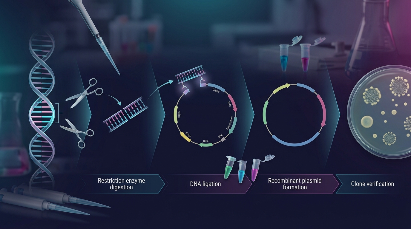
Molecular cloning remains one of the foundational techniques in modern molecular biology, biotechnology, and genetic engineering. At its core lies a deceptively simple pair of tools: restriction enzymes that cut DNA at precise sequences, and DNA ligase that joins fragments back together. This guide explores the biochemistry, methodology, and practical applications of restriction enzyme-based cloning for researchers working with plasmids, recombinant DNA, and protein expression systems.
1. Introduction to Restriction Enzymes
Restriction endonucleases (restriction enzymes) are bacterial proteins that recognize specific short DNA sequences (typically 4–8 base pairs) and cleave the phosphodiester backbone at or near that site. They evolved as part of bacterial restriction-modification (R-M) systems, a primitive immune defense against invading bacteriophage DNA. The bacterium's own DNA is protected by a companion methyltransferase that methylates the same recognition sequence, preventing self-digestion, while unmethylated foreign DNA is cleaved and destroyed.
| Type | Description |
|---|---|
| Type I | Multi-subunit complexes that cut DNA far from their recognition site; rarely used in cloning due to unpredictable cut locations. |
| Type II | The workhorses of molecular biology. They recognize specific palindromic or near-palindromic sequences and cleave within or immediately adjacent to that site, producing reproducible, defined fragments. Nearly all cloning enzymes (EcoRI, BamHI, HindIII, NotI, etc.) belong here. |
| Type III | Cleave DNA a short distance from asymmetric recognition sites; less commonly used. |
| Type IV | Recognize an asymmetric sequence but cut at a defined distance away from it (e.g., BsaI, BsmBI). These are essential for Golden Gate and other scarless cloning strategies. |
2. Recognition Sequences and Cleavage Patterns
Type II restriction enzymes typically recognize palindromic sequences that read the same on both strands in the 5'→3' direction.For example, EcoRI recognizes:
5'-G A A T T C-3'
3'-C T T A A G-5'
Cleavage generates one of three end types:
5' overhangs (sticky ends) : e.g., EcoRI cuts between G and A, leaving a 4-nucleotide 5' overhang (AATT).
3' overhangs (sticky ends) : e.g., PstI generates a 3' overhang.
Blunt ends : e.g., SmaI cuts squarely in the middle of its recognition sequence, leaving no overhang.
Sticky ends are especially valuable in cloning because complementary overhangs allow directional, high-efficiency annealing between insert and vector before ligase seals the nicks. Blunt-end cloning is more universal (any blunt fragment can ligate to any other) but far less efficient and non-directional unless additional strategies are used.
Isoschizomers (enzymes recognizing the same sequence) and neoschizomers (same sequence, different cut position) expand the toolkit for achieving compatible ends between otherwise non-matching enzymes.
3. Star Activity and Enzyme Fidelity
Under non-optimal reaction conditions, restriction enzymes can exhibit star activity, cleaving sequences that resemble but do not exactly match their canonical recognition site.
Common causes include:
Excess glycerol (>5% v/v) in the reaction
High enzyme-to-DNA ratio
Non-optimal pH or ionic strength
Presence of organic solvents (DMSO, ethanol)
Substitution of Mn²⁺ for Mg²⁺
Related products
Sma I Restriction Enzyme
Not I Restriction Enzyme
Xba I Restriction Enzyme
Restriction Enzyme Buffer B, 10X
Spe I Restriction Enzyme
Restriction Enzyme Buffer A, 10X
4. Double Digestion and Buffer Compatibility
Cloning frequently requires cutting a plasmid or insert with two different enzymes simultaneously to create two distinct, non-compatible sticky ends this ensures directional insertion (the fragment can only go in one orientation) and prevents vector self-ligation.

5. Vector Preparation and Dephosphorylation
After linearizing a vector with restriction enzymes, an important optional step is dephosphorylation using alkaline phosphatase (CIP or shrimp alkaline phosphatase, SAP). Removing the 5' phosphate groups from the vector's cut ends prevents self-religation (vector recircularizing without an insert), dramatically improving the ratio of recombinant (insert-containing) colonies to background.
Because ligase requires at least one 5' phosphate to form a phosphodiester bond, a dephosphorylated vector can only ligate with a phosphorylated insert, and the resulting product will contain two nicks (one per strand) that are efficiently repaired by the bacterial host after transformation.
6. Insert Preparation: PCR Products, Gel Purification, and Ligation
Inserts for cloning are commonly generated by PCR using primers engineered with restriction sites at their 5' ends, by digestion of an existing plasmid, or synthetically.
Key steps:

7. Transformation, Selection, and Screening
Ligated products are introduced into chemically competent or electrocompetent bacterial cells (commonly E. coli strains such as DH5α or TOP10). Cells are plated on selective media (antibiotic resistance encoded on the plasmid backbone) to eliminate non-transformed cells.
Screening strategies to identify correctly recombinant clones include:
Blue-white screening -(α-complementation with X-gal/IPTG) when cloning disrupts lacZ.
Colony PCR using primers flanking the insertion site.
Restriction digestion (diagnostic digest) of miniprep plasmid DNA to confirm fragment sizes match predictions.
Sanger sequencing to confirm exact insert sequence, reading frame, and absence of PCR-introduced mutations.
8. Beyond Classical Cloning: Modern Alternatives and Complements
While classical restriction-ligation cloning remains widely used, several complementary or alternative technologies have emerged:
Golden Gate Assembly: Uses Type IIS enzymes (BsaI, BsmBI) whose cut sites lie outside their recognition sequence, allowing scarless, one-pot, multi-fragment assembly with defined junctions.
Gibson Assembly / isothermal assembly: Uses exonuclease, polymerase, and ligase together to join overlapping DNA fragments without restriction sites at all.
Gateway Cloning: Site-specific recombination (att sites) for rapid, restriction-independent shuttling of inserts between vectors.
CRISPR-based cloning/knock-in strategies: Combine restriction/ligation-independent methods with guide RNA-directed genome editing.
Despite these advances, restriction enzyme digestion remains essential for vector linearization, diagnostic verification, and many standard subcloning workflows, and is often combined with these newer methods rather than replaced by them.
9. Applications in Genetic Engineering and Protein Expression
Restriction enzyme-based cloning underlies a vast range of downstream applications

10. Best Practices and Troubleshooting Checklist
A concise checklist for reliable restriction enzyme-based cloning:
Conclusion
Restriction enzymes and molecular cloning form the backbone of recombinant DNA technology, enabling researchers to precisely cut, assemble, and verify custom DNA constructs. Whether building a simple subclone or assembling a multi-fragment expression vector, understanding enzyme biochemistry, buffer compatibility, ligation stoichiometry, and verification strategies is essential for reproducible, efficient molecular biology work.